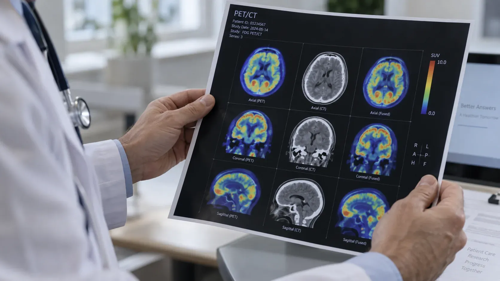
A CT scan evaluates anatomy, detailing the precise size, location, and surrounding structures of a lesion. A PET scan measures metabolic activity, highlighting areas where cells are active, a key insight once cancer has been identified. Because these scans complement each other, they are typically performed simultaneously on a single PET/CT machine to provide a complete diagnostic picture.
It is entirely understandable to question why a second scan is needed when the initial results appeared normal.
Demystifying Your Imaging: How CT and PET Scans Work Together
CT (computed tomography)
Takes X-rays from many angles as the scanner rotates around you, then reconstructs them into cross-sections. Think of it as slicing the body into images a few millimeters apart. It is excellent at anatomy. It can find a mass, measure it precisely, show whether it is pressing on something, and show whether it has grown or shrunk since last time.
What CT cannot do is tell you what a lump is made of. A lymph node enlarged by cancer and a lymph node enlarged by an old infection can look identical.
PET (positron emission tomography)
Works differently. You are injected with a small amount of a radioactive tracer, most often FDG, which is a form of glucose. Cells that are consuming a lot of glucose take up more of it. Many cancers are metabolically busy, so they accumulate more tracer than the tissue around them and show up brighter on the images.
That is the origin of the phrase you will hear people use, that something “lit up.” It means the tracer collected there. It does not, on its own, mean cancer.
Because activity without anatomy is hard to interpret, the two are almost always combined. A PET/CT scanner does both in one sitting and overlays them, so the radiologist can see a bright spot and know exactly which structure it sits in.
Understanding Your Scan Results: What They Can (and Can’t) Reveal
This part is worth understanding carefully, as it’s where most post-scan anxiety usually comes from.
Why PET Scans Aren’t Perfect: Spotting False Positives
FDG is taken up by anything metabolically active, and cancer is not the only thing that qualifies. Infection lights up. Inflammation lights up. So do healing surgical sites, arthritic joints, sarcoidosis, and brown fat, which is a type of tissue in the neck and shoulders that switches on when you are cold and appears on a small percentage of scans. Older series put the false positive rate around 13%.
This is exactly why a bright spot usually leads to a biopsy or a repeat scan rather than straight to a treatment decision.
The Flip Side: When PET Scans Miss Things (False Negatives)
Some cancers simply do not consume much glucose, so they stay dark even when they are there. Slow-growing tumors, certain neuroendocrine tumors, some low-grade lymphomas, and the faint hazy lung nodules called ground-glass opacities are all known for this. In one study of lung adenocarcinomas, none of the pure ground-glass nodules were FDG-avid at all. Very small deposits can also fall below what the scanner can resolve, regardless of how active they are.
A clear PET is genuinely reassuring in most situations. It is not a guarantee, and your team will weigh it against everything else they know.
Understanding Your SUV: Why It’s Not a Simple Cancer Score
If you read your report, you will likely see a number labeled SUV or SUVmax. This simply measures how concentrated the radioactive tracer became in a single spot. Patients often treat this as a “severity score,” but it doesn’t work that way. The number can shift based on your blood sugar, weight, how long you waited after the injection, or even the specific machine used, meaning the same tumor could yield different scores on different days at different hospitals. Radiologists explicitly advise against using any single SUV number as a hard cutoff between benign and malignant. Only compare these numbers with your oncologist, and stick to scans done within the same facility.
Incidental Findings: When Scans Find Things Nobody Was Looking For
Whole-body imaging turns up incidental findings reasonably often: a thyroid nodule, a small adrenal lump, a cyst. Most are harmless. Some need a follow-up scan to confirm that. Being told about one is not the same as being told it is a problem.
Preparing for Your Scan: What Actually Happens on the Day
A CT is quick. You lie on a table that moves through a ring. The scan itself usually takes minutes. You may be given contrast, either to drink or through a vein, which makes blood vessels and organs easier to distinguish. IV contrast contains iodine, so be sure to mention if you have had an allergic reaction before or if you have kidney issues. Because your kidneys process and clear the dye, your team needs to check your kidney function beforehand.
A PET/CT takes longer, and most of that time is waiting rather than scanning. Budget around two hours.
You will be asked to fast beforehand, usually four to six hours, with plain water allowed and encouraged. The point is to keep your blood sugar down, because circulating glucose competes with the tracer. Your blood sugar gets checked on arrival, and if it is too high the scan may be rescheduled. If you have diabetes, ask for specific written instructions well in advance, because the guidance about insulin and meal timing is detailed and getting it wrong wastes the appointment.
Avoid strenuous exercise for a day beforehand, since working muscles take up tracer. Stay warm on the way in, which reduces the brown fat effect. After the injection you rest quietly for roughly an hour, usually in a dim room, without talking or reading much, while the tracer distributes. The scan itself then takes twenty to thirty minutes.
Radiation: What to Expect
These are real doses and worth knowing honestly. The American Cancer Society estimates a CT of the abdomen and pelvis at around 7.7 mSv, roughly what you would get from natural background sources over two and a half years, and a PET/CT at around 22.7 mSv, closer to eight years of background. Most of that comes from the CT component rather than the tracer.
Those numbers sound alarming in isolation and are usually the wrong thing to worry about. The estimated increase in lifetime cancer risk from a scan is very small, and the risk of a cancer going unfound and untreated is not. It is still a fair question to ask whether a particular scan will change the plan.
When Guidelines Vary
Weighing the Benefits: What a PET Scan Adds to Your Treatment Plan
For some cancers and some situations, PET meaningfully changes staging and therefore treatment, particularly for finding disease that has spread beyond where anyone expected. For others it adds cost, time, and radiation without changing what happens next. Guidelines differ by cancer type and by country, and reasonable oncologists reach different conclusions on the same case.
Understanding Follow-Up Imaging: What Surveillance Can (and Can’t) Do
Whether people who have finished treatment should have regular scans to look for recurrence, or be scanned only when symptoms appear, is genuinely contested for several cancers. More scanning does not automatically equate to better care. It can also produce more incidental findings and more anxiety.
The Specific Tracer Used Matters
While FDG is the standard go-to, it isn’t the only option. Disease-specific tracers, like PSMA for prostate cancer or DOTATATE for neuroendocrine tumors, have transformed care for specific conditions. Keep in mind that availability for these specialized tracers can vary widely from one hospital to another.
What to Ask Your Care Team
Four questions that tend to produce useful answers:
- What are you hoping this scan will show, and what would change depending on the result?
- Are we using PET because CT could not answer the question, or in addition to it?
- Is my cancer type one that shows up reliably on FDG PET?
- If something unexpected turns up, what is the plan?
One important step before your scan: let the staff know when booking if you have diabetes, take steroids, or have had recent surgery, an infection, or a biopsy. Each of these can alter how your scan is prepared or interpreted, and they are details the machine can’t detect on its own.
This page explains how these scans work in general terms. It cannot tell you what your own scan shows. Radiology reports are written for clinicians, and SUV values in particular are frequently misread by patients as a measure of how serious a cancer is, which they are not. Bring your report to your oncology team and ask them to walk you through it. If you are waiting on results and finding it hard, most cancer centers have specialist nurses who will talk it through with you before your appointment.
Common questions
Why do I need a PET scan if my CT was normal?
They answer different questions. A CT shows the shape of things: where a lump is, how big, what it sits next to. A PET shows how hard cells in a given spot are working. A lymph node enlarged by cancer and a lymph node enlarged by an old infection can look identical on CT, and a PET can sometimes separate them. In cancer care the two are usually done together on one machine, which is why your paperwork may say PET/CT.
Does something lighting up on a PET scan mean cancer?
No. Lighting up means the tracer collected there, and the tracer is taken up by anything metabolically active. Infection lights up. So does inflammation, healing surgical sites, arthritic joints, sarcoidosis, and brown fat in the neck and shoulders. Older series put the false positive rate around 13%. That is why a bright spot usually leads to a biopsy or a repeat scan rather than straight to a treatment decision.
Can a PET scan miss cancer?
Yes. Some cancers do not consume much glucose and stay dark even when they are there. Slow-growing tumors, certain neuroendocrine tumors, some low-grade lymphomas, and the faint hazy lung nodules called ground-glass opacities are all known for this. Very small deposits can also fall below what the scanner can resolve. A clear PET is genuinely reassuring in most situations, but it is not a guarantee, and your team will weigh it against everything else they know.
What does the SUV number on my report mean?
It is a measure of how concentrated the tracer was in one spot, and it is not a cancer score. The value shifts with your blood sugar, your weight, how long you waited after the injection, and which scanner was used, so the same tumor can produce different numbers on different days in different hospitals. Compare numbers across your own scans only with your oncologist, and not across hospitals at all.
How should I prepare for a PET/CT, and how long does it take?
Budget around two hours, most of it waiting rather than scanning. You will be asked to fast beforehand, usually four to six hours, with plain water allowed and encouraged, because circulating glucose competes with the tracer. Avoid strenuous exercise the day before, and stay warm on the way in. If you have diabetes, ask for specific written instructions well in advance, because the guidance about insulin and meal timing is detailed.
Sources
- Understanding Radiation Risk from Imaging Tests · American Cancer Society, 2026
- Causes and imaging features of false positives and false negatives on 18F-PET/CT in oncologic imaging · Insights into Imaging, 2011
- Limitations and Pitfalls of FDG-PET/CT in Infection and Inflammation · Seminars in Nuclear Medicine, 2021
- Focal FDG uptake in mediastinal brown fat mimicking malignancy: a potential pitfall resolved on PET/CT · American Journal of Roentgenology, 2004
- Adenocarcinoma with BAC Features Presented as the Nonsolid Nodule Is Prone to Be False-Negative on 18F-FDG PET/CT · BioMed Research International, 2015
- Radiation dosimetry of 18F-FDG PET/CT: incorporating exam-specific parameters in dose estimates · BMC Medical Imaging, 2016
- How to Prepare for Your PET/CT Scan - General Directions · Brown University Health, 2024
- PET/MRI Versus PET/CT for Whole-Body Staging: Results from a Single-Center Observational Study on 1,003 Sequential Examinations · Journal of Nuclear Medicine, 2020