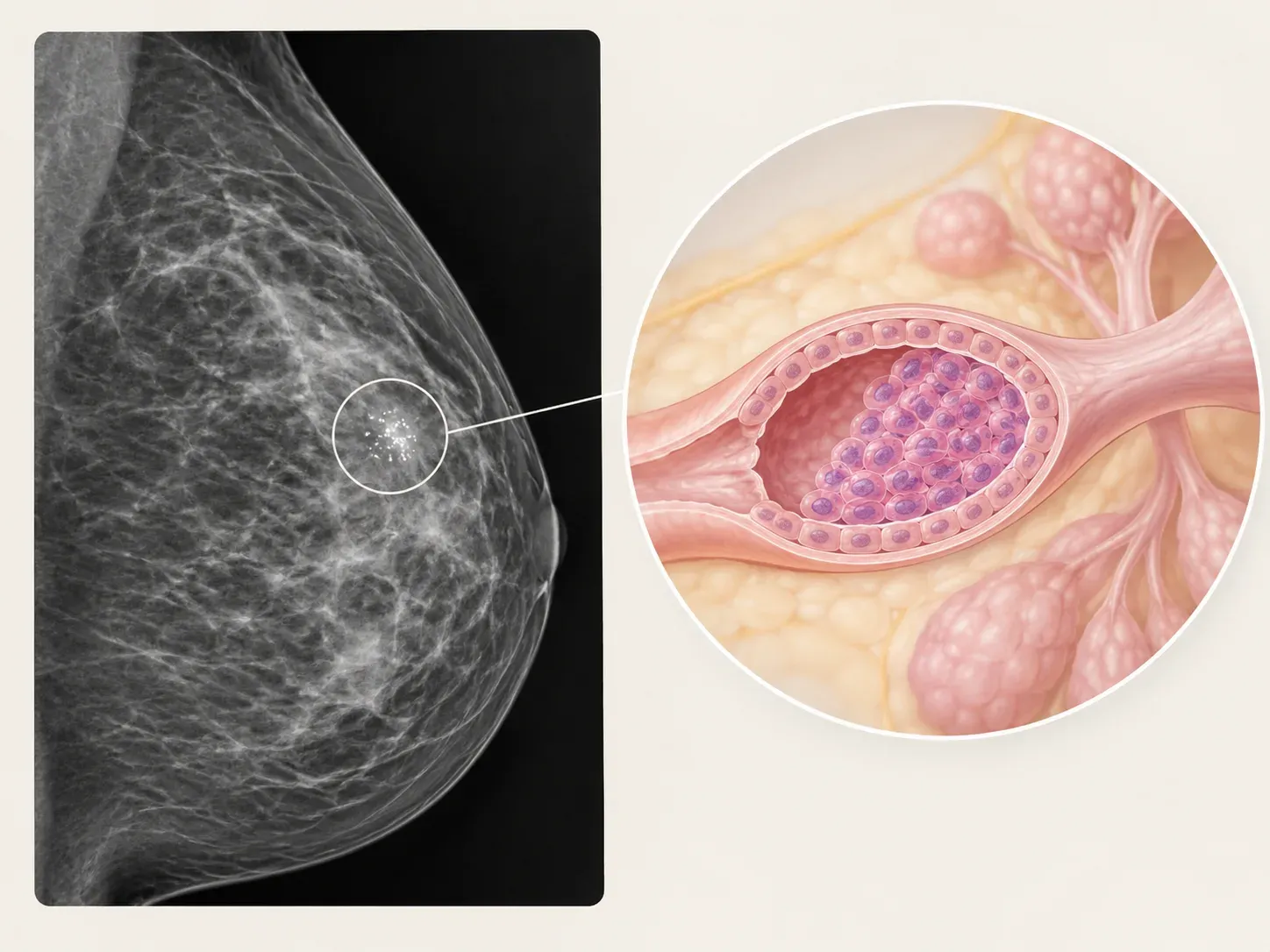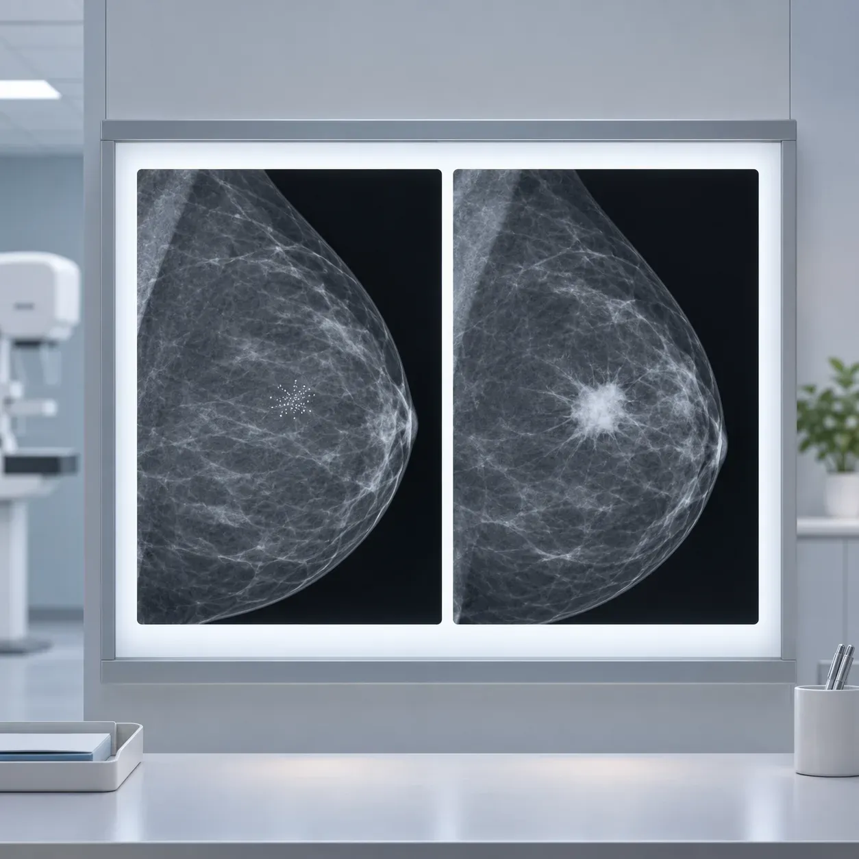
The entire distinction comes down to one layer of cells.
Breast ducts are lined on the outside by a thin boundary made of myoepithelial cells and a basement membrane. In DCIS, abnormal cells are multiplying inside the duct and that boundary is still intact. In IDC, cells have broken through it into the surrounding breast tissue, where they can reach blood vessels and lymph channels.
That single structural difference is why DCIS is stage 0 and cannot spread elsewhere in the body, while IDC is staged, can involve lymph nodes, and may need treatment beyond the breast. This guide covers what actually changes between the two, and the three situations that confuse people most: reports listing both, diagnoses that change after surgery, and the category that sits in between.
If you want the fuller picture of DCIS itself, including survival data and the debate about overtreatment, that’s covered in our guide to what DCIS is.
One Layer of Cells: How Pathologists Tell Them Apart
Invasive ductal carcinoma, increasingly written in reports as invasive breast carcinoma of no special type, is the most common form of invasive breast cancer, accounting for about 80% of cases. The word invasive means only that the cells have crossed the duct boundary. It does not mean advanced, aggressive, or spread.
The diagnosis rests on finding, or failing to find, that boundary layer. When a pathologist can’t tell from the appearance alone, they apply special stains that light up myoepithelial cells specifically, using markers such as p63 and smooth muscle myosin heavy chain. If an intact rim of those cells surrounds the abnormal glands, it’s in situ. If the rim is gone and tumor cells sit loose in the surrounding tissue, it’s invasive.
This is worth knowing because it explains why the distinction is sometimes described as uncertain on a small sample, and why your report may mention additional stains. For more on how these reports are structured, see our guide to reading a pathology report.
What Actually Changes in Treatment
Staging. DCIS is always stage 0. IDC is staged from I to IV depending on size, lymph nodes, and whether it has spread.
Lymph nodes. This is the clearest practical difference. Because DCIS can’t reach the lymph nodes, sentinel node biopsy generally isn’t performed alongside a lumpectomy for pure DCIS, though it’s usually done with a mastectomy for technical reasons. For IDC, assessing the nodes is standard.
Systemic treatment. DCIS is not treated with chemotherapy or HER2-targeted drugs. For IDC, both may be on the table depending on the tumor’s biology, size, nodes, and receptor status.
Genomic testing. Tests that estimate recurrence risk and the likely benefit of chemotherapy are routine in many invasive cancers. For DCIS, genomic testing exists but is used differently, mainly to inform decisions about radiation.
HER2. Tested routinely in IDC because it directs treatment. Not standard practice for DCIS, because there’s no HER2-targeted therapy for it.
Endocrine therapy. Offered for both when the cells are hormone receptor positive, though the purpose differs. In DCIS it reduces the risk of a future cancer. In IDC it also treats the cancer that’s there.
Surgery. Here the two are similar. Both are usually treated with lumpectomy followed by radiation, or with mastectomy, chosen on much the same grounds.
Prognosis. Twenty-year breast cancer mortality after DCIS is around 3%. For IDC it depends almost entirely on stage and biology, and spans a very wide range.
When Your Report Says Both
This is the most common scenario of all, and the one that causes the most unnecessary alarm. Most invasive ductal carcinomas have DCIS sitting alongside them, which makes biological sense, since the invasive cancer usually arose from it.
The invasive component drives everything. Your stage, your treatment plan, and your prognosis are determined by the IDC. The DCIS around it does not add to the severity.
Your tumor size is the invasive part only. This is the single most useful thing on this page, because people routinely misread it. Staging protocols are explicit: the measurement includes only the area of invasive carcinoma and excludes the surrounding DCIS. So if your report describes 4 cm of DCIS with a 7 mm invasive focus, your tumor is 7 mm, not 4 cm. Those are very different conversations, and the difference matters enormously.
One term you may see. An extensive intraductal component usually means DCIS makes up at least a quarter of the area within the invasive tumor and also extends beyond it. It can affect surgical planning, because clear margins may be harder to achieve, but it isn’t a measure of how dangerous the cancer is.
When the Diagnosis Changes After Surgery
Some people are told they have DCIS after a needle biopsy, then told after surgery that invasive cancer was found. This is distressing and it is not a mistake.
A core biopsy samples a tiny fraction of the lesion. It takes a few narrow cores from an area that may span centimeters. If a small invasive focus sits somewhere the needle didn’t reach, it won’t appear until the whole thing is removed and examined.
How often does it happen? Reported rates vary widely depending on the population and how the biopsy was done, from under 10% to over 40%, with many series landing somewhere around 15% to 25%. A large study developing a prediction tool found 14.1% in its main group and 10.7% in external validation. Among people with the kind of low-risk DCIS eligible for active monitoring trials, upstaging rates were lower, roughly 6% to 10%.
What makes it more likely: a larger lesion, a mass visible on imaging rather than calcifications alone, high-grade DCIS, and a biopsy that reported possible invasion.
What happens next. The plan is revised to treat invasive cancer. The most common practical consequence is needing a second procedure to assess the lymph nodes, since that wasn’t done the first time. It may also open up systemic treatment that wasn’t relevant before.
Worth saying plainly: this usually means a small invasive cancer was found early, during an operation you were already having. It is a change in plan rather than a disaster.
Microinvasion: The Category In Between
Occasionally a report describes DCIS with microinvasion, meaning one or more tiny areas where cells have crossed the boundary, with no single focus larger than 1 mm. It’s staged as T1mi, and it accounts for less than 1% of all breast cancers.
Technically this is invasive cancer, so it sits on the IDC side of the line. In practice it behaves much more like DCIS: overall survival is about the same, though the risk of recurrence is somewhat higher. Lymph node assessment is usually considered, because more invasion is sometimes found once everything is examined.
If this is what your report says, the honest summary is that you are in a genuinely intermediate category, and management varies more between centers than it does for either pure DCIS or straightforward IDC.
Your Next Steps
Questions worth asking:
- “Is my diagnosis pure DCIS, invasive cancer, or both?”
- “If both, what’s the size of the invasive part specifically?”
- “Was a sentinel node biopsy done or planned, and why or why not?”
- “Given my biopsy findings, what’s the chance invasion is found at surgery?”
- “Will the plan change if the final pathology differs from the biopsy?”
Two things to keep in mind.
Your final pathology is the one that counts. A core biopsy result is a strong indication, not a verdict. Decisions about systemic treatment generally wait for the surgical specimen.
Ask for a copy of both reports and keep them together. The distinction between in situ and invasive, and the size of the invasive component, are the two numbers that drive nearly everything that follows, and you will need them for second opinions and trial screening.
A final word. Finding out that your diagnosis crossed from in situ to invasive is a genuinely significant change, and it’s reasonable to feel that. It’s also worth keeping the scale in view: most invasive ductal carcinoma found this way is small, node-negative, and caught at a point where treatment works well. One layer of cells changes the treatment plan considerably. It changes the outlook far less than the word invasive makes it sound.
This page explains the distinction between in situ and invasive ductal carcinoma in general terms. It cannot interpret your own report, and terminology varies between laboratories. Upstaging rates quoted here differ substantially between studies depending on biopsy technique, lesion size, and the population studied, so they should not be read as your personal risk. Staging, surgical planning, and decisions about lymph node assessment and systemic treatment belong with your own breast surgical and oncology team.
Common questions
What is the difference between DCIS and IDC?
In DCIS, abnormal cells are multiplying inside the breast duct and the boundary of myoepithelial cells and basement membrane around it is still intact. In IDC, cells have broken through that boundary into the surrounding breast tissue, where they can reach blood vessels and lymph channels. That single structural difference is why DCIS is stage 0 and cannot spread elsewhere in the body, while IDC is staged, can involve lymph nodes, and may need treatment beyond the breast.
If my pathology report mentions both DCIS and IDC, which one matters?
The invasive component drives everything. Your stage, your treatment plan, and your prognosis are determined by the IDC, and the DCIS around it does not add to the severity. Your tumor size is the invasive part only, so if your report describes 4 cm of DCIS with a 7 mm invasive focus, your tumor is 7 mm, not 4 cm.
Why did my diagnosis change from DCIS to invasive cancer after surgery?
A core biopsy samples a tiny fraction of the lesion, so a small invasive focus the needle didn't reach won't appear until the whole thing is removed and examined. This is distressing, but it is not a mistake. Reported rates vary widely, from under 10% to over 40%, and it is more likely with a larger lesion, a mass visible on imaging, high-grade DCIS, or a biopsy that reported possible invasion.
Do I need a sentinel lymph node biopsy for DCIS?
Because DCIS can't reach the lymph nodes, sentinel node biopsy generally isn't performed alongside a lumpectomy for pure DCIS, though it's usually done with a mastectomy for technical reasons. For IDC, assessing the nodes is standard. If invasive cancer is found at surgery, the most common practical consequence is needing a second procedure to assess the lymph nodes.
What does DCIS with microinvasion mean?
It means one or more tiny areas where cells have crossed the duct boundary, with no single focus larger than 1 mm. It's staged as T1mi and is technically invasive cancer, but in practice it behaves much more like DCIS: overall survival is about the same, though the risk of recurrence is somewhat higher. Lymph node assessment is usually considered, because more invasion is sometimes found once everything is examined.
Sources
- Protocol for the Examination of Specimens From Patients With Invasive Carcinoma of the Breast · College of American Pathologists, 2018
- Invasive Ductal Carcinoma (IDC) · Breastcancer.org, 2024
- A Validated Nomogram to Predict Upstaging of Ductal Carcinoma in Situ to Invasive Disease · Annals of Surgical Oncology, 2017 · doi:10.1245/s10434-017-5927-y
- Predicting upstaging of DCIS to invasive disease: radiologist’s predictive performance · Academic Radiology, 2020 · doi:10.1016/j.acra.2019.12.009
- Pathological underestimation and biomarkers concordance rates in breast cancer patients diagnosed with ductal carcinoma in situ at preoperative biopsy · Scientific Reports, 2022 · doi:10.1038/s41598-022-06206-7
- Microinvasive · PathologyOutlines.com, 2024
- Microinvasive Carcinoma versus Ductal Carcinoma In Situ: A Comparison of Clinicopathological Features and Clinical Outcomes · Journal of Breast Cancer, 2018 · doi:10.4048/jbc.2018.21.2.197
- Prognostic significance of microinvasion with ductal carcinoma in situ of the breast: a meta-analysis · Breast Cancer Research and Treatment, 2023 · doi:10.1007/s10549-022-06800-3
- Axillary staging in ductal carcinoma in situ with microinvasion: A meta-analysis · Surgical Oncology, 2021 · doi:10.1016/j.suronc.2021.101557
- Association of a Diagnosis of Ductal Carcinoma In Situ With Death From Breast Cancer · JAMA Network Open, 2020 · doi:10.1001/jamanetworkopen.2020.17124
- Surgical Upstaging Rates for Vacuum Assisted Biopsy Proven DCIS: Implications for Active Surveillance Trials · Annals of Surgical Oncology, 2017 · doi:10.1245/s10434-017-6018-9